Because every image counts
Philips ultrasound solutions connect technology, clinicians and patients to empower patient-focused care and elevate the healthcare experience. Our comprehensive portfolio – ranging from ultra-mobile handheld devices to premium cart-based systems – is designed to empower clinical confidence, with innovations that enhance imaging accuracy and performance. We’re committed to designing sustainable solutions for reliability, scalability and ease-of-use. Paired with shared architecture and comprehensive service programs, our solutions deliver lifetime value to you and your patients.

The new Flash Ultrasound System 5100 POC - built for the way you work at the point of care


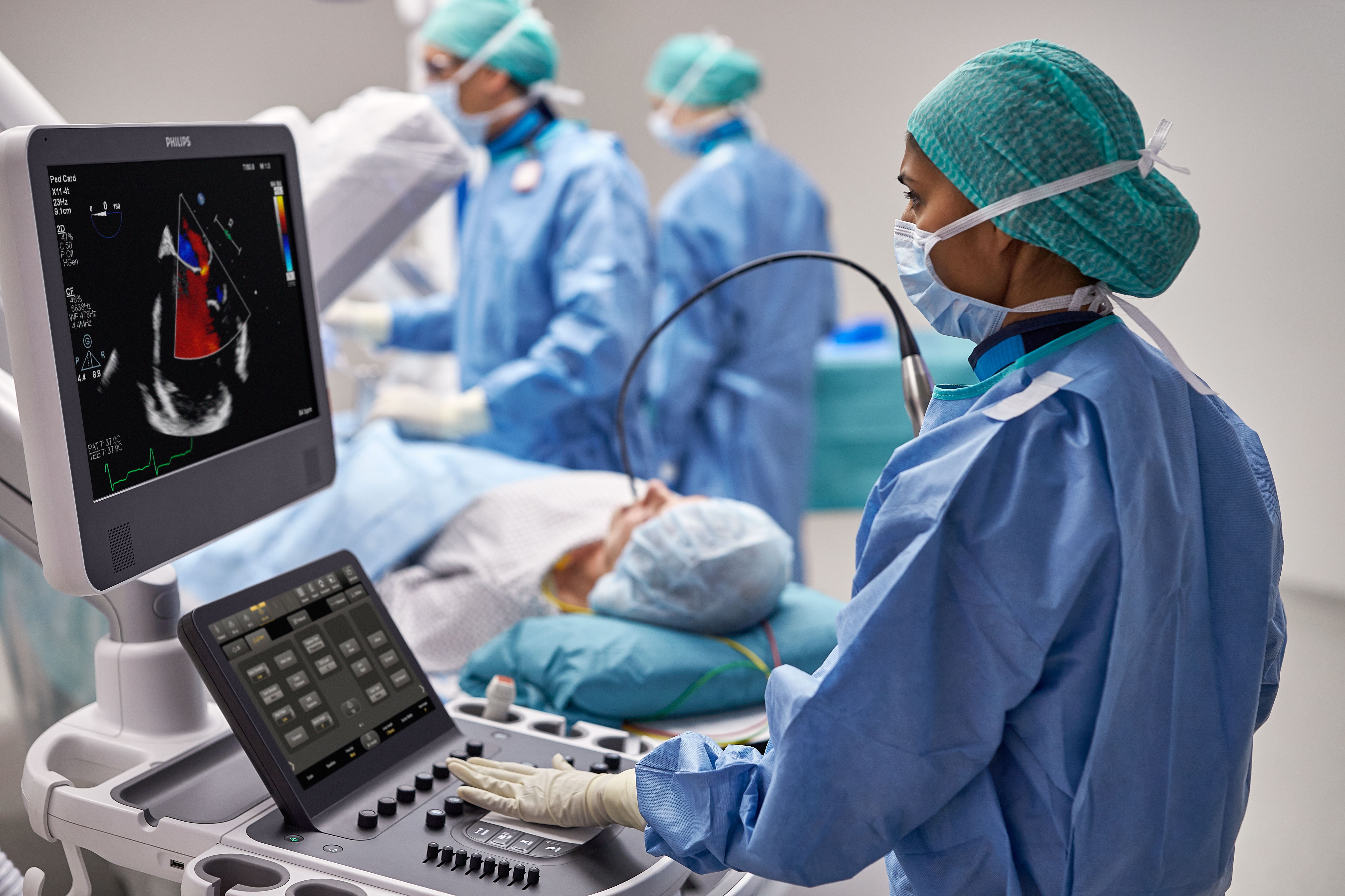
Philips AI-based ultrasound solutions integrate into everyday clinical workflows and augment user skills.

Auto ElastQ improves liver elastography precision, reproducibility and ease of use by providing assistance with frame selection and ROI location.

xMATRIX transducer technology enhances image clarity and eases workflow to make exams faster and easier for both clinicians and patients.

Available on Philips ultrasound machines, Flow Viewer defines vasculature with a 3D-like appearance and reduced flash artifact.

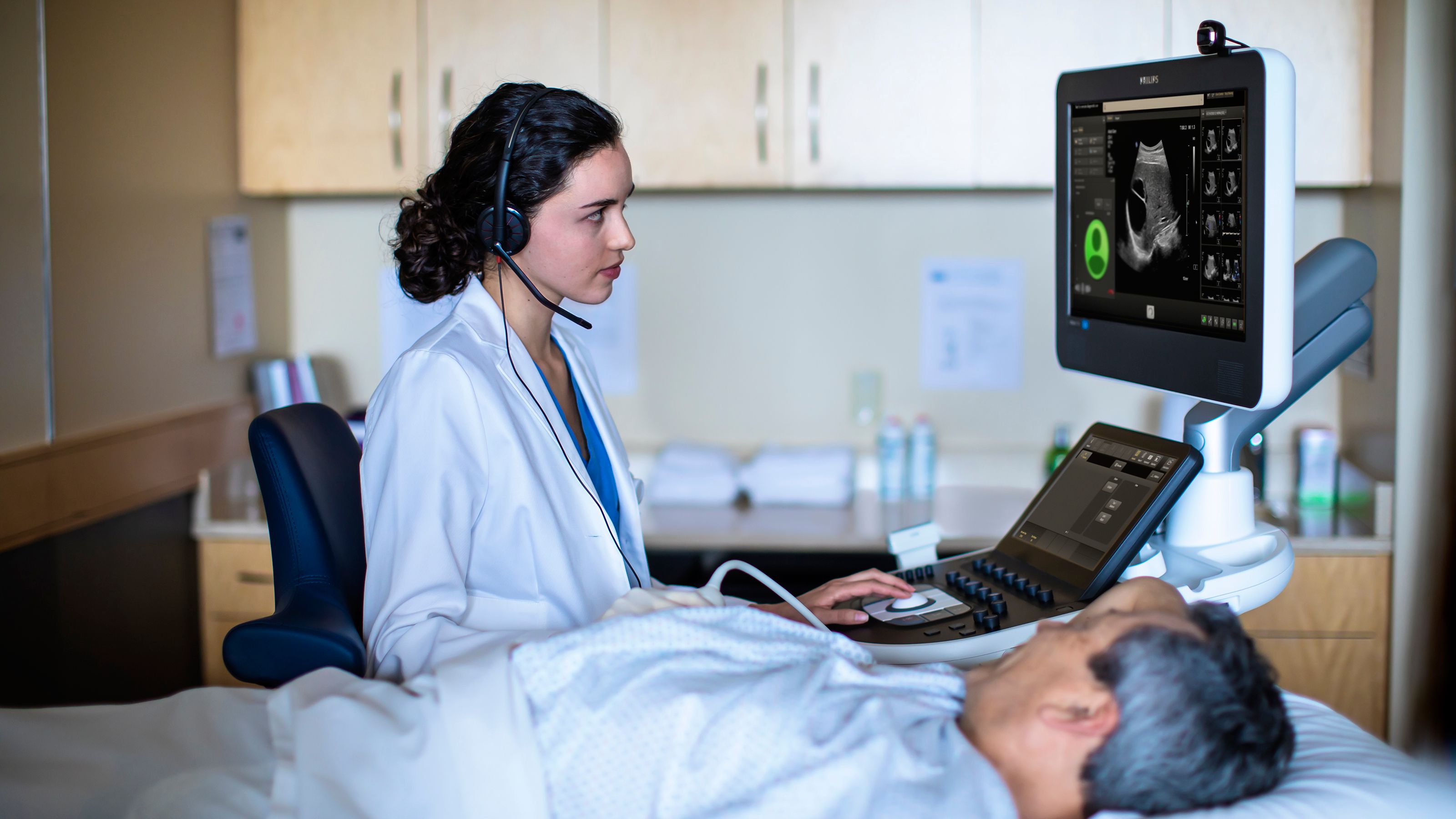
Give patients access to your team’s full expertise, regardless of location. Learn more.


