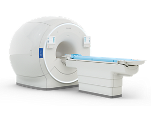
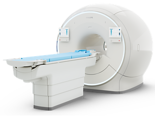
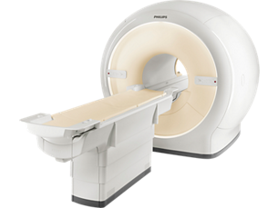
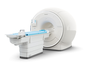
MR 7700
Experience breakthrough innovation in 3.0T imaging with the unique design of the Philips MR 7700 imaging system, enhanced with XP gradients and artificial intelligence (AI). The system is built to address a pressing need to deliver on the clinical expectations of today, and to facilitate the most demanding research programs.
The MR 7700 provides high accuracy, power, and endurance to support confident diagnosis for every patient. It is the system of choice for highest quality diffusion imaging and advanced neuroscience.
Extend your scanning capabilities with a fully integrated multi-nuclei imaging and spectroscopy solution to explore new clinical pathways without sacrificing clinical imaging workflow or wide-bore patient comfort.
What’s more? The MR 7700 promises a great experience for both users and patients through the ease-of-use features of a well-designed clinical 3.0T scanner together with a no compromise workflow. Now scientists and clinicians alike can schedule without conflict.
View product


