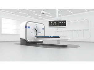
CT Brain Perfusion

Determine areas of reduced cerebral blood flow compared to the contralateral hemisphere​

Find similar products

Generates qualitative and quantitative information about image intensity changes over time. It supports identification of hypo-perfused areas in acute stroke by use of quantitative color maps. The perfusion and summary maps can be automatically generated and sent to PACS for convenient reviewing.​ And sent to a pre-defined list of recipients via email in less than 2 mins(4)(5) . Quality indicators (‘traffic lights’) highlighting potential acquisition inaccuracies that may affect results reliability. With studies of sufficient scan duration, permeability analysis can be used as an assessment of the contrast agent permeation of the blood-brain barrier. The application offers automatic detection of reference artery, vein, mirror line and brain mask as well as 3D motion correction.​

  • Enhanced in ISP 12.1.6 (2) Configurable settings now allow to select either TTP or Tmax for calculating the summary maps. (3) Summary maps are calculated according to default thresholds. Enable user to configure calculated parameters. (4) Content sent via email is not for diagnostic use.

You are about to visit a Philips global content page

Continue

You are about to visit a Philips global content page

Continue

Our site can best be viewed with the latest version of Microsoft Edge, Google Chrome or Firefox.

Вие влизате в българския уебсайт на Philips Healthcare.
Please select the checkbox

Уебсайтът е предназначен само за хора, които използват медицински изделия като професионалисти, хора, действащи за здравни организации или организации, търгуващи по занятие с медицински изделия.

Вие сте човек, който отговаря на горните критерии ?