MRCAT Head and Neck

MR-RT clinical application

Find similar products

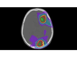
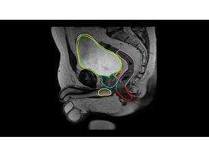
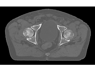
MRCAT Head and Neck enables the use of MRI as a primary imaging modality for radiotherapy planning of patients with soft tissue tumors in the Head and Neck, without the need for CT. Powered by AI, MRCAT (MR for Calculating ATtenuation) images with CT-like density information are automatically generated from a single, high-resolution MR scan and can be used for CT-equivalent¹ dose calculations and accurate² patient positioning.

Features
MR Simulation
Unleash the real power of MR simulation
Unleash the real power of MR simulation

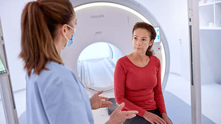
MRCAT Head and Neck lets you plan radiation therapy using MRI only, without the need for CT. Within just one fast MR exam, MRCAT Head and Neck provides excellent soft-tissue contrast for target and OAR delineation, and CT-like density information for dose calculations. This not only extends the benefits of MRI’s excellent soft-tissue contrast to radiotherapy planning and adaptive strategies, but it also eliminates arduous, error-prone CT-MRI registration from the process.

Unleash the real power of MR simulation

Unleash the real power of MR simulation
MRCAT Head and Neck lets you plan radiation therapy using MRI only, without the need for CT. Within just one fast MR exam, MRCAT Head and Neck provides excellent soft-tissue contrast for target and OAR delineation, and CT-like density information for dose calculations. This not only extends the benefits of MRI’s excellent soft-tissue contrast to radiotherapy planning and adaptive strategies, but it also eliminates arduous, error-prone CT-MRI registration from the process.

Unleash the real power of MR simulation

MRCAT Head and Neck lets you plan radiation therapy using MRI only, without the need for CT. Within just one fast MR exam, MRCAT Head and Neck provides excellent soft-tissue contrast for target and OAR delineation, and CT-like density information for dose calculations. This not only extends the benefits of MRI’s excellent soft-tissue contrast to radiotherapy planning and adaptive strategies, but it also eliminates arduous, error-prone CT-MRI registration from the process.
Click here for more information
MR Simulation
Unleash the real power of MR simulation

Unleash the real power of MR simulation

MRCAT Head and Neck lets you plan radiation therapy using MRI only, without the need for CT. Within just one fast MR exam, MRCAT Head and Neck provides excellent soft-tissue contrast for target and OAR delineation, and CT-like density information for dose calculations. This not only extends the benefits of MRI’s excellent soft-tissue contrast to radiotherapy planning and adaptive strategies, but it also eliminates arduous, error-prone CT-MRI registration from the process.
Protocol
Consistent, accurate imaging protocol
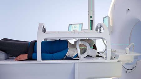
Consistent, accurate imaging protocol

Image acquisition is made easy by a dedicated ExamCard that includes a single, multi-contrast 3D T1W mDIXON scan, which is standardized to deliver consistent results and high geometric accuracy. The MRCAT Head and Neck scan can be acquired before or after contrast agent administration. Additional sequences, e.g. for delineation, can be easily added to the protocol to address your specific clinical needs.

Consistent, accurate imaging protocol

Consistent, accurate imaging protocol
Image acquisition is made easy by a dedicated ExamCard that includes a single, multi-contrast 3D T1W mDIXON scan, which is standardized to deliver consistent results and high geometric accuracy. The MRCAT Head and Neck scan can be acquired before or after contrast agent administration. Additional sequences, e.g. for delineation, can be easily added to the protocol to address your specific clinical needs.

Consistent, accurate imaging protocol

Image acquisition is made easy by a dedicated ExamCard that includes a single, multi-contrast 3D T1W mDIXON scan, which is standardized to deliver consistent results and high geometric accuracy. The MRCAT Head and Neck scan can be acquired before or after contrast agent administration. Additional sequences, e.g. for delineation, can be easily added to the protocol to address your specific clinical needs.
Click here for more information
Protocol
Consistent, accurate imaging protocol

Consistent, accurate imaging protocol

Image acquisition is made easy by a dedicated ExamCard that includes a single, multi-contrast 3D T1W mDIXON scan, which is standardized to deliver consistent results and high geometric accuracy. The MRCAT Head and Neck scan can be acquired before or after contrast agent administration. Additional sequences, e.g. for delineation, can be easily added to the protocol to address your specific clinical needs.
Patient Comfort
Short scan times promote patient comfort
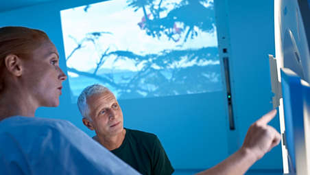
Short scan times promote patient comfort

Head and neck cancer patients often find it difficult to endure an MRI exam, especially when immobilized by a thermoplastic mask. The high-resolution 3D MRCAT source scan is accelerated by Compressed SENSE and requires < 3 minutes to complete – while covering a large FOV (up to 380 mm in the feet-head direction). This promotes patient comfort, as well as productivity by minimizing time in the scanner.

Short scan times promote patient comfort

Short scan times promote patient comfort
Head and neck cancer patients often find it difficult to endure an MRI exam, especially when immobilized by a thermoplastic mask. The high-resolution 3D MRCAT source scan is accelerated by Compressed SENSE and requires < 3 minutes to complete – while covering a large FOV (up to 380 mm in the feet-head direction). This promotes patient comfort, as well as productivity by minimizing time in the scanner.

Short scan times promote patient comfort

Head and neck cancer patients often find it difficult to endure an MRI exam, especially when immobilized by a thermoplastic mask. The high-resolution 3D MRCAT source scan is accelerated by Compressed SENSE and requires < 3 minutes to complete – while covering a large FOV (up to 380 mm in the feet-head direction). This promotes patient comfort, as well as productivity by minimizing time in the scanner.
Click here for more information
Patient Comfort
Short scan times promote patient comfort

Short scan times promote patient comfort

Head and neck cancer patients often find it difficult to endure an MRI exam, especially when immobilized by a thermoplastic mask. The high-resolution 3D MRCAT source scan is accelerated by Compressed SENSE and requires < 3 minutes to complete – while covering a large FOV (up to 380 mm in the feet-head direction). This promotes patient comfort, as well as productivity by minimizing time in the scanner.
Synthetic CT images using AI
Automatic generation of synthetic CT images using AI
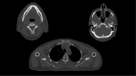
Automatic generation of synthetic CT images using AI

MRCAT Head and Neck uses Artificial Intelligence (AI) for fast computation of MRCAT attenuation maps based on the mDIXON source scan. The resulting MRCAT density maps provide continuous Hounsfield Units for CT-like image appearance and are shown right at the MR console for immediate review.

Automatic generation of synthetic CT images using AI

Automatic generation of synthetic CT images using AI
MRCAT Head and Neck uses Artificial Intelligence (AI) for fast computation of MRCAT attenuation maps based on the mDIXON source scan. The resulting MRCAT density maps provide continuous Hounsfield Units for CT-like image appearance and are shown right at the MR console for immediate review.

Automatic generation of synthetic CT images using AI

MRCAT Head and Neck uses Artificial Intelligence (AI) for fast computation of MRCAT attenuation maps based on the mDIXON source scan. The resulting MRCAT density maps provide continuous Hounsfield Units for CT-like image appearance and are shown right at the MR console for immediate review.
Click here for more information
Synthetic CT images using AI
Automatic generation of synthetic CT images using AI

Automatic generation of synthetic CT images using AI

MRCAT Head and Neck uses Artificial Intelligence (AI) for fast computation of MRCAT attenuation maps based on the mDIXON source scan. The resulting MRCAT density maps provide continuous Hounsfield Units for CT-like image appearance and are shown right at the MR console for immediate review.
Accuracy in dose planning
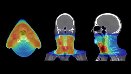
Accuracy in dose planning

MRCAT Head and Neck has been designed with strict radiotherapy accuracy requirements in mind. MRCAT image acquisition is geometrically accurate³ and MRCAT-based dose plans are as robust and accurate¹ as CT-based plans, promoting confidence in dose planning.

Accuracy in dose planning

Accuracy in dose planning
MRCAT Head and Neck has been designed with strict radiotherapy accuracy requirements in mind. MRCAT image acquisition is geometrically accurate³ and MRCAT-based dose plans are as robust and accurate¹ as CT-based plans, promoting confidence in dose planning.

Accuracy in dose planning

MRCAT Head and Neck has been designed with strict radiotherapy accuracy requirements in mind. MRCAT image acquisition is geometrically accurate³ and MRCAT-based dose plans are as robust and accurate¹ as CT-based plans, promoting confidence in dose planning.
Click here for more information
Accuracy in dose planning

Accuracy in dose planning

MRCAT Head and Neck has been designed with strict radiotherapy accuracy requirements in mind. MRCAT image acquisition is geometrically accurate³ and MRCAT-based dose plans are as robust and accurate¹ as CT-based plans, promoting confidence in dose planning.
MR-CAT patient positioning
Rely on MRCAT-based patient positioning
Rely on MRCAT-based patient positioning

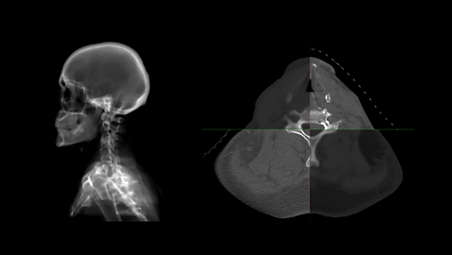
You can use MRCAT data for patient positioning verification at the linac that is as accurate² as with conventional CT images. MRCAT’s continuous Hounsfield units, with the look and feel of CT, enable CBCT-based positioning using soft-tissue or bony contrast. You can also use MRCAT data to generate MR-based digitally reconstructed radiographs (DRRs) to allow for patient positioning using bony structures.

Rely on MRCAT-based patient positioning

Rely on MRCAT-based patient positioning
You can use MRCAT data for patient positioning verification at the linac that is as accurate² as with conventional CT images. MRCAT’s continuous Hounsfield units, with the look and feel of CT, enable CBCT-based positioning using soft-tissue or bony contrast. You can also use MRCAT data to generate MR-based digitally reconstructed radiographs (DRRs) to allow for patient positioning using bony structures.

Rely on MRCAT-based patient positioning

You can use MRCAT data for patient positioning verification at the linac that is as accurate² as with conventional CT images. MRCAT’s continuous Hounsfield units, with the look and feel of CT, enable CBCT-based positioning using soft-tissue or bony contrast. You can also use MRCAT data to generate MR-based digitally reconstructed radiographs (DRRs) to allow for patient positioning using bony structures.
Click here for more information
MR-CAT patient positioning
Rely on MRCAT-based patient positioning

Rely on MRCAT-based patient positioning

You can use MRCAT data for patient positioning verification at the linac that is as accurate² as with conventional CT images. MRCAT’s continuous Hounsfield units, with the look and feel of CT, enable CBCT-based positioning using soft-tissue or bony contrast. You can also use MRCAT data to generate MR-based digitally reconstructed radiographs (DRRs) to allow for patient positioning using bony structures.
MR-Only radiotherapy
Put Philips MR-only radiotherapy to work today

Put Philips MR-only radiotherapy to work today

To successfully bring MR-only radiotherapy planning into your clinical routine, we recognize that you must look beyond the imaging itself and address important steps such as patient marking, position verification, and quality assurance. We are prepared to support you throughout this process. To this end, we offer dedicated workflow descriptions, best practice sharing and tailored training support, designed to assist as you adopt this new treatment paradigm.

Put Philips MR-only radiotherapy to work today

Put Philips MR-only radiotherapy to work today
To successfully bring MR-only radiotherapy planning into your clinical routine, we recognize that you must look beyond the imaging itself and address important steps such as patient marking, position verification, and quality assurance. We are prepared to support you throughout this process. To this end, we offer dedicated workflow descriptions, best practice sharing and tailored training support, designed to assist as you adopt this new treatment paradigm.

Put Philips MR-only radiotherapy to work today

To successfully bring MR-only radiotherapy planning into your clinical routine, we recognize that you must look beyond the imaging itself and address important steps such as patient marking, position verification, and quality assurance. We are prepared to support you throughout this process. To this end, we offer dedicated workflow descriptions, best practice sharing and tailored training support, designed to assist as you adopt this new treatment paradigm.
Click here for more information
MR-Only radiotherapy
Put Philips MR-only radiotherapy to work today

Put Philips MR-only radiotherapy to work today

To successfully bring MR-only radiotherapy planning into your clinical routine, we recognize that you must look beyond the imaging itself and address important steps such as patient marking, position verification, and quality assurance. We are prepared to support you throughout this process. To this end, we offer dedicated workflow descriptions, best practice sharing and tailored training support, designed to assist as you adopt this new treatment paradigm.
  • MR Simulation
  • Protocol
  • Patient Comfort
  • Synthetic CT images using AI
See all features
MR Simulation
Unleash the real power of MR simulation

Unleash the real power of MR simulation

MRCAT Head and Neck lets you plan radiation therapy using MRI only, without the need for CT. Within just one fast MR exam, MRCAT Head and Neck provides excellent soft-tissue contrast for target and OAR delineation, and CT-like density information for dose calculations. This not only extends the benefits of MRI’s excellent soft-tissue contrast to radiotherapy planning and adaptive strategies, but it also eliminates arduous, error-prone CT-MRI registration from the process.

Unleash the real power of MR simulation

Unleash the real power of MR simulation
MRCAT Head and Neck lets you plan radiation therapy using MRI only, without the need for CT. Within just one fast MR exam, MRCAT Head and Neck provides excellent soft-tissue contrast for target and OAR delineation, and CT-like density information for dose calculations. This not only extends the benefits of MRI’s excellent soft-tissue contrast to radiotherapy planning and adaptive strategies, but it also eliminates arduous, error-prone CT-MRI registration from the process.

Unleash the real power of MR simulation

MRCAT Head and Neck lets you plan radiation therapy using MRI only, without the need for CT. Within just one fast MR exam, MRCAT Head and Neck provides excellent soft-tissue contrast for target and OAR delineation, and CT-like density information for dose calculations. This not only extends the benefits of MRI’s excellent soft-tissue contrast to radiotherapy planning and adaptive strategies, but it also eliminates arduous, error-prone CT-MRI registration from the process.
Click here for more information
MR Simulation
Unleash the real power of MR simulation

Unleash the real power of MR simulation

MRCAT Head and Neck lets you plan radiation therapy using MRI only, without the need for CT. Within just one fast MR exam, MRCAT Head and Neck provides excellent soft-tissue contrast for target and OAR delineation, and CT-like density information for dose calculations. This not only extends the benefits of MRI’s excellent soft-tissue contrast to radiotherapy planning and adaptive strategies, but it also eliminates arduous, error-prone CT-MRI registration from the process.
Protocol
Consistent, accurate imaging protocol

Consistent, accurate imaging protocol

Image acquisition is made easy by a dedicated ExamCard that includes a single, multi-contrast 3D T1W mDIXON scan, which is standardized to deliver consistent results and high geometric accuracy. The MRCAT Head and Neck scan can be acquired before or after contrast agent administration. Additional sequences, e.g. for delineation, can be easily added to the protocol to address your specific clinical needs.

Consistent, accurate imaging protocol

Consistent, accurate imaging protocol
Image acquisition is made easy by a dedicated ExamCard that includes a single, multi-contrast 3D T1W mDIXON scan, which is standardized to deliver consistent results and high geometric accuracy. The MRCAT Head and Neck scan can be acquired before or after contrast agent administration. Additional sequences, e.g. for delineation, can be easily added to the protocol to address your specific clinical needs.

Consistent, accurate imaging protocol

Image acquisition is made easy by a dedicated ExamCard that includes a single, multi-contrast 3D T1W mDIXON scan, which is standardized to deliver consistent results and high geometric accuracy. The MRCAT Head and Neck scan can be acquired before or after contrast agent administration. Additional sequences, e.g. for delineation, can be easily added to the protocol to address your specific clinical needs.
Click here for more information
Protocol
Consistent, accurate imaging protocol

Consistent, accurate imaging protocol

Image acquisition is made easy by a dedicated ExamCard that includes a single, multi-contrast 3D T1W mDIXON scan, which is standardized to deliver consistent results and high geometric accuracy. The MRCAT Head and Neck scan can be acquired before or after contrast agent administration. Additional sequences, e.g. for delineation, can be easily added to the protocol to address your specific clinical needs.
Patient Comfort
Short scan times promote patient comfort

Short scan times promote patient comfort

Head and neck cancer patients often find it difficult to endure an MRI exam, especially when immobilized by a thermoplastic mask. The high-resolution 3D MRCAT source scan is accelerated by Compressed SENSE and requires < 3 minutes to complete – while covering a large FOV (up to 380 mm in the feet-head direction). This promotes patient comfort, as well as productivity by minimizing time in the scanner.

Short scan times promote patient comfort

Short scan times promote patient comfort
Head and neck cancer patients often find it difficult to endure an MRI exam, especially when immobilized by a thermoplastic mask. The high-resolution 3D MRCAT source scan is accelerated by Compressed SENSE and requires < 3 minutes to complete – while covering a large FOV (up to 380 mm in the feet-head direction). This promotes patient comfort, as well as productivity by minimizing time in the scanner.

Short scan times promote patient comfort

Head and neck cancer patients often find it difficult to endure an MRI exam, especially when immobilized by a thermoplastic mask. The high-resolution 3D MRCAT source scan is accelerated by Compressed SENSE and requires < 3 minutes to complete – while covering a large FOV (up to 380 mm in the feet-head direction). This promotes patient comfort, as well as productivity by minimizing time in the scanner.
Click here for more information
Patient Comfort
Short scan times promote patient comfort

Short scan times promote patient comfort

Head and neck cancer patients often find it difficult to endure an MRI exam, especially when immobilized by a thermoplastic mask. The high-resolution 3D MRCAT source scan is accelerated by Compressed SENSE and requires < 3 minutes to complete – while covering a large FOV (up to 380 mm in the feet-head direction). This promotes patient comfort, as well as productivity by minimizing time in the scanner.
Synthetic CT images using AI
Automatic generation of synthetic CT images using AI

Automatic generation of synthetic CT images using AI

MRCAT Head and Neck uses Artificial Intelligence (AI) for fast computation of MRCAT attenuation maps based on the mDIXON source scan. The resulting MRCAT density maps provide continuous Hounsfield Units for CT-like image appearance and are shown right at the MR console for immediate review.

Automatic generation of synthetic CT images using AI

Automatic generation of synthetic CT images using AI
MRCAT Head and Neck uses Artificial Intelligence (AI) for fast computation of MRCAT attenuation maps based on the mDIXON source scan. The resulting MRCAT density maps provide continuous Hounsfield Units for CT-like image appearance and are shown right at the MR console for immediate review.

Automatic generation of synthetic CT images using AI

MRCAT Head and Neck uses Artificial Intelligence (AI) for fast computation of MRCAT attenuation maps based on the mDIXON source scan. The resulting MRCAT density maps provide continuous Hounsfield Units for CT-like image appearance and are shown right at the MR console for immediate review.
Click here for more information
Synthetic CT images using AI
Automatic generation of synthetic CT images using AI

Automatic generation of synthetic CT images using AI

MRCAT Head and Neck uses Artificial Intelligence (AI) for fast computation of MRCAT attenuation maps based on the mDIXON source scan. The resulting MRCAT density maps provide continuous Hounsfield Units for CT-like image appearance and are shown right at the MR console for immediate review.
Accuracy in dose planning

Accuracy in dose planning

MRCAT Head and Neck has been designed with strict radiotherapy accuracy requirements in mind. MRCAT image acquisition is geometrically accurate³ and MRCAT-based dose plans are as robust and accurate¹ as CT-based plans, promoting confidence in dose planning.

Accuracy in dose planning

Accuracy in dose planning
MRCAT Head and Neck has been designed with strict radiotherapy accuracy requirements in mind. MRCAT image acquisition is geometrically accurate³ and MRCAT-based dose plans are as robust and accurate¹ as CT-based plans, promoting confidence in dose planning.

Accuracy in dose planning

MRCAT Head and Neck has been designed with strict radiotherapy accuracy requirements in mind. MRCAT image acquisition is geometrically accurate³ and MRCAT-based dose plans are as robust and accurate¹ as CT-based plans, promoting confidence in dose planning.
Click here for more information
Accuracy in dose planning

Accuracy in dose planning

MRCAT Head and Neck has been designed with strict radiotherapy accuracy requirements in mind. MRCAT image acquisition is geometrically accurate³ and MRCAT-based dose plans are as robust and accurate¹ as CT-based plans, promoting confidence in dose planning.
MR-CAT patient positioning
Rely on MRCAT-based patient positioning

Rely on MRCAT-based patient positioning

You can use MRCAT data for patient positioning verification at the linac that is as accurate² as with conventional CT images. MRCAT’s continuous Hounsfield units, with the look and feel of CT, enable CBCT-based positioning using soft-tissue or bony contrast. You can also use MRCAT data to generate MR-based digitally reconstructed radiographs (DRRs) to allow for patient positioning using bony structures.

Rely on MRCAT-based patient positioning

Rely on MRCAT-based patient positioning
You can use MRCAT data for patient positioning verification at the linac that is as accurate² as with conventional CT images. MRCAT’s continuous Hounsfield units, with the look and feel of CT, enable CBCT-based positioning using soft-tissue or bony contrast. You can also use MRCAT data to generate MR-based digitally reconstructed radiographs (DRRs) to allow for patient positioning using bony structures.

Rely on MRCAT-based patient positioning

You can use MRCAT data for patient positioning verification at the linac that is as accurate² as with conventional CT images. MRCAT’s continuous Hounsfield units, with the look and feel of CT, enable CBCT-based positioning using soft-tissue or bony contrast. You can also use MRCAT data to generate MR-based digitally reconstructed radiographs (DRRs) to allow for patient positioning using bony structures.
Click here for more information
MR-CAT patient positioning
Rely on MRCAT-based patient positioning

Rely on MRCAT-based patient positioning

You can use MRCAT data for patient positioning verification at the linac that is as accurate² as with conventional CT images. MRCAT’s continuous Hounsfield units, with the look and feel of CT, enable CBCT-based positioning using soft-tissue or bony contrast. You can also use MRCAT data to generate MR-based digitally reconstructed radiographs (DRRs) to allow for patient positioning using bony structures.
MR-Only radiotherapy
Put Philips MR-only radiotherapy to work today

Put Philips MR-only radiotherapy to work today

To successfully bring MR-only radiotherapy planning into your clinical routine, we recognize that you must look beyond the imaging itself and address important steps such as patient marking, position verification, and quality assurance. We are prepared to support you throughout this process. To this end, we offer dedicated workflow descriptions, best practice sharing and tailored training support, designed to assist as you adopt this new treatment paradigm.

Put Philips MR-only radiotherapy to work today

Put Philips MR-only radiotherapy to work today
To successfully bring MR-only radiotherapy planning into your clinical routine, we recognize that you must look beyond the imaging itself and address important steps such as patient marking, position verification, and quality assurance. We are prepared to support you throughout this process. To this end, we offer dedicated workflow descriptions, best practice sharing and tailored training support, designed to assist as you adopt this new treatment paradigm.

Put Philips MR-only radiotherapy to work today

To successfully bring MR-only radiotherapy planning into your clinical routine, we recognize that you must look beyond the imaging itself and address important steps such as patient marking, position verification, and quality assurance. We are prepared to support you throughout this process. To this end, we offer dedicated workflow descriptions, best practice sharing and tailored training support, designed to assist as you adopt this new treatment paradigm.
Click here for more information
MR-Only radiotherapy
Put Philips MR-only radiotherapy to work today

Put Philips MR-only radiotherapy to work today

To successfully bring MR-only radiotherapy planning into your clinical routine, we recognize that you must look beyond the imaging itself and address important steps such as patient marking, position verification, and quality assurance. We are prepared to support you throughout this process. To this end, we offer dedicated workflow descriptions, best practice sharing and tailored training support, designed to assist as you adopt this new treatment paradigm.

Specifications

MRCAT Head and Neck
MRCAT Head and Neck
Compatibility MR system
  • Ingenia MR-RT 1.5T and 3.0T
  • Ingenia Ambition MR-RT 1.5T S and X
  • Ingenia Elition MR-RT 3.0T S and X
  • Ingenia MR-RT Evolution 1.5T and 3.0T
MRCAT Head and Neck
MRCAT Head and Neck
Compatibility MR system
  • Ingenia MR-RT 1.5T and 3.0T
  • Ingenia Ambition MR-RT 1.5T S and X
  • Ingenia Elition MR-RT 3.0T S and X
  • Ingenia MR-RT Evolution 1.5T and 3.0T
See all specifications
MRCAT Head and Neck
MRCAT Head and Neck
Compatibility MR system
  • Ingenia MR-RT 1.5T and 3.0T
  • Ingenia Ambition MR-RT 1.5T S and X
  • Ingenia Elition MR-RT 3.0T S and X
  • Ingenia MR-RT Evolution 1.5T and 3.0T
  • ¹ The mean dose in the PTV does not differ more than 1% in MRCAT-based plans as compared to CT-based plans for 95% of the patient cases
  • ² Accurate means: MRCAT-based patient positioning is within 2 mm accuracy for bony anatomy compared to CT-based patient positioning for 95% of cases
  • ³ Accurate means: MRCAT provides < ± 1 mm total system geometric accuracy of image data in < 20 cm Diameter Spherical Volume (DSV) and < ± 2 mm total system geometric accuracy of image data in < 40 cm Diameter Spherical Volume (DSV)* * Limited to 32 cm in z-direction in more than 95% of the points within the volume
  • MRCAT Head and Neck is not available for sale in all markets.
You are about to visit a Philips global content page

Continue

You are about to visit a Philips global content page

Continue

Our site can best be viewed with the latest version of Microsoft Edge, Google Chrome or Firefox.

Вие влизате в българския уебсайт на Philips Healthcare.
Please select the checkbox

Уебсайтът е предназначен само за хора, които използват медицински изделия като професионалисти, хора, действащи за здравни организации или организации, търгуващи по занятие с медицински изделия.

Вие сте човек, който отговаря на горните критерии ?