FieldStrength MRI magazine
User experiences - June 2015
After introducing the latest methods, radiologists get more information in the same time, making them more confident in their diagnoses.
Since the installation of Ingenia 1.5T in the new building of Meander Medical Center, the MRI team has enjoyed many successes in improving their MRI scanning by implementing the latest techniques available to them, such as Diffusion TSE for distortion-free images, mDIXON TSE for adding fat-free imaging without adding time, and motion correction with MultiVane XD.
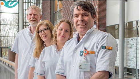
From left to right: MRI technologists Gerrit Kooiker, Jane van Straelen, Natasja van Bree, radiologist Ben Heggelman, MD Ben Heggelman, MD, was trained as a radiologist at the Erasmus University Hospital Rotterdam. He has worked for 27 years at Meander Medical Center.
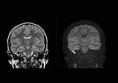
“We have high sensitivity and specificity for cholesteatoma acquisition with Diffusion TSE.”
Gradual transformation to a new standard
Obtaining high quality MR imaging in the head and neck area can be quite demanding, because the susceptibility changes at the many (curved) interfaces between air, tissue and bone may lower the quality of fat suppression, in diffusion weighted imaging in particular. In addition, the area is prone to motion, which also affects image quality. Driven by the desire for artifact-free imaging that facilitates easy and confident diagnoses, radiologist Ben Heggelman, MD, and his team have implemented some of the latest techniques. Because of the heavy patient load at the department the changes were gradual to allow for finetuning the sequences to their preferences. This process went together with a growing diagnostic confidence among the radiologists. After a few months, the team is passionate about the improvements they achieved.
Excellent cholesteatoma imaging with Diffusion TSE
“Imaging cholesteatoma, benign tumors of the middle ear, has been a huge challenge,” says Dr. Heggelman. “We used to do CT, but then we were unsure if we were looking at an inflammation or a cholesteatoma. Also determining if residual cholesteatoma exist after surgery or visualizing recurrence used to be very difficult. Adding Diffusion TSE in our MRI protocol now effectively addresses this.” “Diffusion TSE is far less sensitive to susceptibility differences than previously used EPI sequences. We appreciate the high resolution and the robustness of the sequence. The quality is so good that our confidence has increased. Also our ENT (ear, nose, throat) physicians are excited about the high resolution, the excellent lesion delineation and the sensitivity and specificity.”

Meander Medical Center (Amersfoort, the Netherlands) is a 600-bed community hospital. Since last year the hospital is concentrated in a beautiful new building, with only single-patient rooms, on the outskirts of Amersfoort. The Radiology Department employs 13 radiologists, 3 nuclear medicine physicians and in total 13 residents. The department operates three 1.5T Philips MRI systems, including Ingenia 1.5T and Achieva 1.5T dStream. (www.meandermc.nl)
“With mDIXON, we not only get T2 images but we also get a T2 with fat suppression ‘for free’ in the same scan.”
mDIXON TSE boosts homogeneity and efficiency
Dr. Heggelman raves about mDIXON TSE because it provides him an extra image series without having to add another scan. “With mDIXON TSE, we not only get a T2-weighted series, but we also get the T2 fat suppressed images ‘for free’ in the same scan. I feel much more confident with the homogeneous fat suppression that mDIXON TSE provides under virtually all conditions, even in this challenging anatomy. SPAIR and SPIR weren’t good enough due to the susceptibility problems in the air cavities, so that fat suppression was not homogeneous over the whole field of view. That made it difficult to see whether something was enhancing or the fat suppression was not good enough.”
“To me the most remarkable fact is that mDIXON TSE provides us T2-weighted images with and without fat suppression at the same time. In the past we needed two separate sequences for that, so it does save some time.”
Value of mDIXON TSE image quality
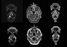
“The excellent image quality of mDIXON helps us a lot. We can, for instance, see the foramina in the skull base very well. Also our confidence in imaging of the facial nerve and the trigeminal nerve is highly improved. Visualizing these nerves properly used to be difficult because they run very close to the air cavities. However, it is very important to know if there are abnormalities or not. I’m very satisfied with the possibilities of mDIXON TSE.”
“These methods save time and produce beautiful images.”
mDIXON TSE in MRI of orbits
mDIXON TSE also benefits MR imaging of orbits, according to Dr. Heggelman. “Using mDIXON TSE helps us reduce fat suppression problems due to susceptibility. The mDIXON TSE orbital images look outstanding. So, also here I get excellent fat suppressed images, and on top of that, the in-phase images as well in the same time.” “We can also use mDIXON TSE for post-contrast imaging and choose to have T1-weighted both with and without fat suppression at the same time. In the past, it took us two scans to get the same information!”
Motion reduction
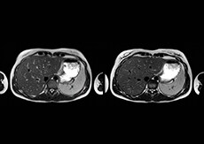
“We also love MultiVane XD for motion reduction in imaging. We find this a huge step forward. We use it in the head, and of course in the upper abdomen, and the images are outstanding most of the time. And it can be combined with dS SENSE parallel imaging for speed.” “We have compared image quality of FLAIR with MultiVane XD versus FLAIR without MultiVane XD. In 15 of the 40 patients studied, we saw motion artifacts on plain FLAIR brain images. The FLAIR images with MultiVane XD were motion-free in 39 of 40 patients and showed slight motion artifacts in only one patient.”
More information without extending time slot
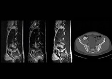
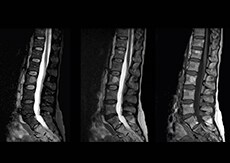
“In our lumbar spine MRI, the value of mDIXON TSE is so obvious. Normally we perform T1 and T2 scans in sagittal and transverse orientation.It used to take too much time to add a sagittal T2 with good fat suppression.But now, using mDIXON TSE, we get the sagittal T2 fat suppressed images ‘for free’, that is: without adding time.” “Diagnostically that is a great benefit. I sometimes see abnormalities in the fat suppressed sagittal T2 that would be quite challenging to notice in the T2 without fat suppression. There have been several diagnoses that I could make easier because of our exam setup with mDIXON TSE, such as sacrum insufficiency fractures and sacroileitis; these were more challenging with our previous exam setup.”
A boost for efficiency
“The described techniques have taken us a big step forward,” concludes Dr. Heggelman. “Especially in more challenging regions, these methods save time and produce beautiful images. I feel much more confident with the high image quality and fewer artifacts.”
*Premium IQ is defined as image quality compared to analog Achieva Results from case studies are not predictive of results in other cases. Results in other cases may vary. Results obtained by facilities described in this issue may not be typical for all facilities. Images that are not part of User experiences articles and that are not labeled otherwise are created by Philips.
Related information
More from FieldStrength
